Dr. Andrew Van Praagh, Lab Manager at Bruker Biospin
Written December 12, 2017 by Imanis Life Sciences, Rochester MN
Welcome to the series “A Vision for the Future,” where Imanis Life Sciences highlights new technologies that are accelerating scientific progress in noninvasive imaging. This episode features Dr. Andrew Van Praagh, Lab Manager at the Preclinical Imaging Lab at Bruker Biospin. Bruker is a leading company in the development of small animal imaging instruments that are simplifying the approach to in vivo experimentation while allowing for the responsible use of experimental animals, two outcomes that Imanis strongly supports in our work.
In this conversation, Andrew delves into:
- exciting additions to the Bruker product portfolio that are advancing noninvasive imaging
- the benefits of a multimodal system
- the importance of using high-quality imaging reagents
- current challenges facing noninvasive imaging
- opportunities for synergies in optical and nuclear imaging.
Watch the video of our conversation, or continue to read the article.
Sophisticated New Technologies
In 2015, Bruker made an exciting addition to their product portfolio: the In Vivo Xtreme II system. The machine boasts five imaging modalities.
Andrew elaborates, “It provides three distinct optical imaging modalities: fluorescence, luminescence, and reflectance, and additionally, high-resolution digital 2d X-Ray, and direct radionuclide imaging.
This sophisticated system allows the user to uncover both anatomical and functional information with the help of:
- flexible multimodal bioluminescent imaging
- Cherenkov radiation imaging
- fluorescent imaging
- direct radioisotopic imaging (DRI)
- X-Ray imaging
Read more about the assets of the In Vivo Xtreme II in this application note from Bruker.
The Power of Multimodality
Why does having five imaging modalities make the In Vivo Xtreme II system so powerful for scientists? Andrew explains “when you have five imaging modalities…you can create more nuanced and powerful animal model systems.”
He illuminates the power of multimodality with an example from a specific study.
“Let’s say you’re doing an oncology model, and you want to evaluate the biodistribution and size of your tumor masses. You can do that with the reporter gene constructs in the cell lines produced by Imanis. If you wanted to make use of a Lung Adenocarcinoma cell line, knowing they produce both fluorescence and luminescence, you could in one experimental template use the firefly luciferase expression of these cells to detect the location in vivo by luminescence.”
“Then, let’s say you wanted to evaluate a therapeutic – not only for its efficacy but for its overall Pharmacokinetic/Pharmacodynamic (PK/PD) – you can do they by tagging that therapeutic with a fluorescent probe. In your animal model then, you’d do sequential luminescent and fluorescent imaging, probably followed up by X-Ray imaging. You could then do trimodal registration of those data to determine the location of the tumor, the location of the therapeutic, all with regard to the anatomical structure of the mouse.”
Importance of Imaging Reagent Quality
Even if one has access to the most sophisticated equipment with multimodal imaging capabilities, imaging reagent quality is paramount to achieving superior results. As Andrew says, “The quality of imaging reagents is essentially the difference between data and no data.”
Andrew and his team often run in vitro serial dilution of various cell lines to look at the flux rate of each and decide which cell line to go with. He tested the Murine Melanoma and Lung Adenocarcinoma cell line from Imanis and found them to be noticeably stronger in constitutive Luciferase expression.
This made the study even more exciting for the Bruker team, as they were able to use their powerful equipment to its full potential with the assistance of quality imaging reagents. In Andrew’s words, “It was the quality of the Imanis cell lines in regards to Luciferase activity that really made the study more interesting.”
Challenges Facing Noninvasive Imaging
In the midst of exciting developments in noninvasive imaging, scientists in the field are also working to overcome several challenges. Andrew identifies some of the key obstacles he has encountered or observed.
“The largest challenge has been providing a multimodal architecture for preclinical noninvasive imaging that includes both the optical modalities and the anatomical modalities. When we’re looking at the ability to combine optical and PET with X-Ray or CT for example, that has always been a challenge. Bruker is pushing the envelope by developing PET-MR systems that will bring together both the metabolic data provided by PET and the anatomical framework provided by MR.”
Synergies for Optical and Nuclear
Combining nuclear and optical imaging provides significant opportunities to advance current imaging techniques. Andrew discusses some of these opportunities, and how he is working to realize their great potential.
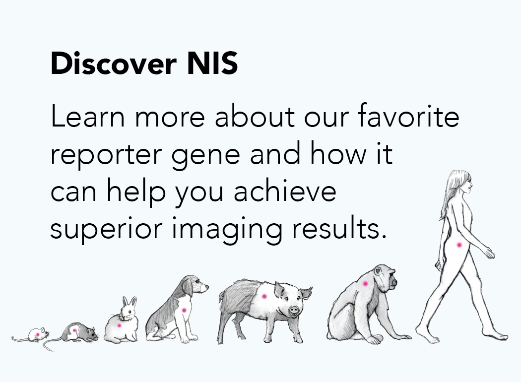
“The potential for synergies are really unlimited. This can be expanded when you use nuclear kinetic reporters like the Sodium Iodide Symporter that are expressed in various Imanis cell lines. This is the future. This is the area that I personally, at the Bruker Import Facility Lab, will be exploring: the use of the convergence of optical and nuclear.”
Thank you to Dr. Andrew Van Praagh for contributing his perspective gained from many years of working with noninvasive imaging, and thank you for watching or reading.
—
This is episode 3 of our “Vision for the Future” series where we examine how industry leaders are progressing noninvasive in vivo imaging.
Explore our second episode with Andrew Heinmiller, Product Manager at FujiFilm Visualsonics here.
Explore our first episode with Dr. Freek Beekman, founder of MI labs, here.