HT1080-Fluc-Neo/eGFP-Puro is a polyclonal population of the human fibrosarcoma cell line HT1080 (ATCC® CCL-121™). To achieve stable reporter expression in the polyclonal population, parental HT1080 cells were transduced with LV-Fluc-P2A-Neo (LV011) and LV-eGFP-PGK-Puro (LV031) and selected using G418 and puromycin. LV-Fluc-P2A-Neo encodes the firefly luciferase (Fluc) cDNA linked to the neomycin resistance gene (Neo) via a P2A cleavage peptide under the spleen focus-forming virus (SFFV) promoter. LV-eGFP-PGK-Puro encodes the enhanced green fluorescent protein (eGFP) cDNA under the SFFV promoter and the puromycin resistance gene (Puro) under the phosphoglycerate kinase promoter (PGK).
*The ATCC trademark and trade name and any and all ATCC catalog numbers are trademarks of the American Type Culture Collection.
This cell line has been tested for mycoplasma contamination and is certified mycoplasma free.
The parental HT1080 cell line has been authenticated and certified free of interspecies cross contamination by short tandem repeat (STR) profiling with 9 STR loci.
 Low and high density cell morphology (200x)
 HT1080-Fluc-Neo/eGFP-Puro (green) or control (HT1080-Fluc-Puro) cells were fixed with paraformaldehyde and analyzed by flow cytometry (20,000 events).
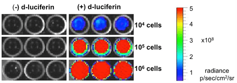 104, 105, or 106 cells were placed in wells of a 96-well plate and 0.3 mg of D-luciferin was added to the indicated wells. The plate was immediately imaged using a Xenogen IVIS Spectrum.
 HT1080-Fluc-Puro cells were injected subcutaneously into the right flanks of female NCR athymic mice. Mice were imaged at day 0 on the day of cell implantation and subsequently at days 7 and 16 using a Perkin Elmer IVIS® Spectrum system, at 10-15 minutes post intraperitoneal injection of D-luciferin at 150 mg/kg. Tumor size was measured using calipers. Data from a representative mouse (ID#6329) is shown.
Complete Growth Medium: DMEM supplemented with 10% FBS, 1X Penicillin/Streptomycin, 1.25 mg/mL G418, and 1 µg/mL puromycin.
The addition of G418 and puromycin to the complete growth medium maintains high dual reporter expression over continued passage of the cells. It is highly recommended, especially if the cells undergo multiple passages prior to being used for studies.
These cells should be grown in the indicated medium and passaged when they reach confluency. For routine passaging, cells are recommended to be split at a 1:10 ratio every 3-4 days.
These cells are suitable for in vitro and in vivo experimentation.
HT1080 cells are a xenograph model for fibrosarcoma and form primary tumors and distant metastases (lung, liver, and brain) post implantation into immunosuppressed mice.1,2
The Fluc transgene facilitates in vivo noninvasive bioluminescent imaging of implanted cells. eGFP is not recommended for whole animal in-live imaging. Rather, samples can be collected postmortem for analysis by conventional fluorescence microscopy or flow cytometry.
The cells can be amplified in vitro and used to generate additional frozen stocks. Cryopreservation of low passage stocks is recommended. Frozen stocks should be preserved in a designated cryopreservation medium.
These cells were generated via lentiviral vector transduction. The lentiviral vector used for transduction was a self-inactivating (SIN) vector in which the viral enhancer and promoter have been deleted. Transcription inactivation of the LTR in the SIN provirus increases biosafety by preventing mobilization by replication competent viruses and enables regulated expression of the genes from the internal promoters without cis-acting effects of the LTR3. Nevertheless, all work with these cells should be performed under biosafety-level 2 (BSL2) conditions by trained personnel. Institutional requirements may permit handling of these cells under BSL1 conditions if certain criteria are met.
References:
1Rasheed et al. Cancer. 1974. 33:1027-1033.
2Praus et al. Gene Therapy. 1999. 6:227-236.
3Miyoshi et al. J Virol 1998. 72:8150-8157.

Don’t see what you need in our catalog? Our scientific team engineers cell lines, viral vectors, and assays and provides the characterization data you need to move forward with confidence.