The Nalm6-Fluc-Puro/HuCD19-Puro (low) cell line stably expresses firefly luciferase and levels of human CD19 that are well-below the endogenous levels normally observed in Nalm6 cells (see Characterization tab for data). The cells are designed for in vitro and in vivo studies of tumor cells expressing CD19. These cells can be used in combination with other Imanis Nalm6-Fluc-Puro cell lines that are part of the CD19 panel comprised of Nalm6-Fluc-Puro cells with various CD19 surface receptor densities:
To generate this cell line, Nalm6-Fluc-Puro/CD19-KO cells were transduced with a human CD19-encoding lentivirus. A low HuCD19 expressing population was generated by selection using a methylcellulose based semi-solid medium.
This cell line has been tested for mycoplasma contamination and is certified mycoplasma free.
The parental Nalm6 cell line was licensed and purchased directly from ATCC*.
*The ATCC trademark and trade name and any and all ATCC catalog numbers are trademarks of the American Type Culture Collection.
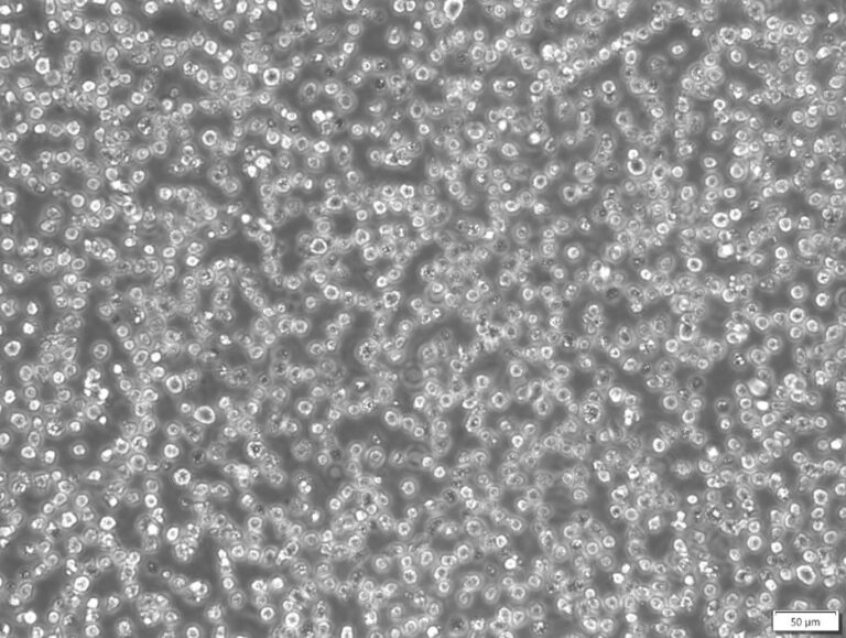 Phase cell photo taken at 200x magnification
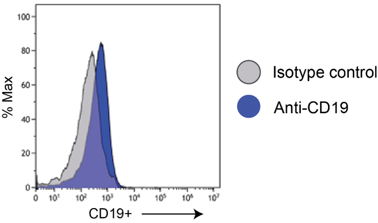
Nalm6-Fluc-Puro/HuCD19-Puro (low) cells were stained with an anti-CD19 (blue) or isotype control (grey) antibody and analyzed by flow cytometry.
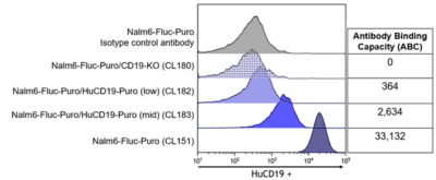
The indicated cell lines were stained with an anti-CD19 (blue) or isotype control (grey) antibody and analyzed by flow cytometry. The antibody binding capacity, representing the number of CD19 receptors on the surface of the cells was quantitated using a Quantum-Simply cellular kit.
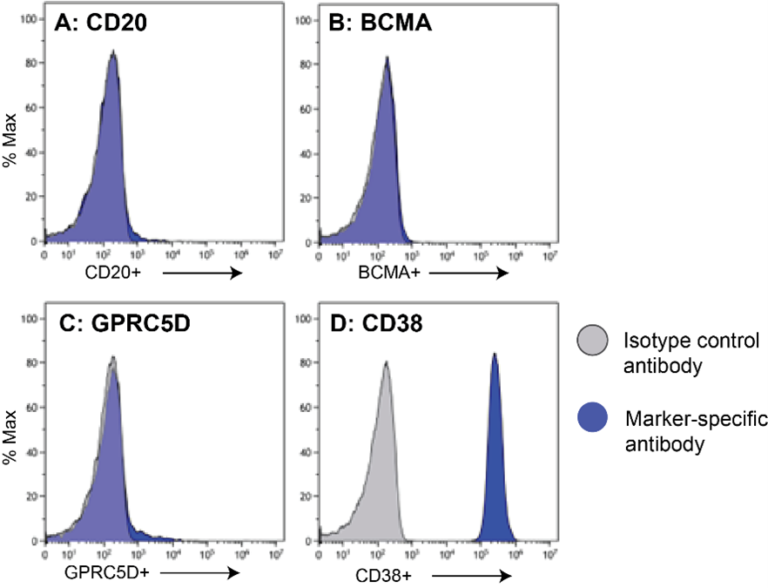 Nalm6-Fluc-Puro/HuCD19-Puro (low)cells (blue) were stained with anti-CD20 (A), anti-BCMA (B), anti-GPRC5D (C), anti-CD38 (D), or isotype control (grey; all panels) antibody and analyzed by flow cytometry.
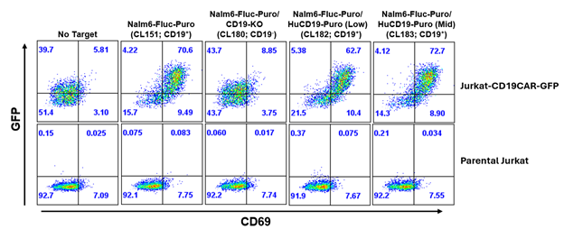 Jurkat T cells expressing a CD19-CAR and GFP were generated by lentivector transduction. Tranduced or untransduced parental Jurkat were incubated in a 1:1 ratio with the indicated Nalm6 cell lines and the following day flow cytometry was performed. The CD3+ Jurkat cells were analyzed for CD19CAR-GFP expression and CD69 expression, which is upregulated in activated T cells.
Complete Growth Medium: RPMI-1640 supplemented with 10% fetal bovine serum (FBS), 10 mM HEPES, and 1% Penicillin/Streptomycin.
For long-term maintenance (more than two to three weeks) addition of 1 µg/mL of puromycin to the media is recommended to maintain high Fluc and HuCD19 expression.
These cells should be grown in the indicated medium and subcultured as needed to maintain a density between 5 x 10⁵ and 3 x 10⁶ cells/mL. The cells can be passaged by dilution in fresh medium, with occasional passaging using centrifugation to limit the amount of debris in cultures.
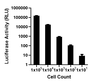
These cells are suitable for in vitro and in vivo experimentation. The Fluc transgene facilitates in vivo noninvasive bioluminescent imaging of implanted cells and quantitation of cells in vitro.
These cells were generated via lentiviral vector transduction. The lentiviral vectors used for transduction were self-inactivating (SIN) vectors in which the viral enhancer and promoter have been deleted. Transcription inactivation of the LTR in the SIN provirus increases biosafety by preventing mobilization by replication competent viruses and enables regulated expression of the genes from the internal promoters without cis-acting effects of the LTR1. Nevertheless, all work with these cells should be performed under biosafety-level 2 (BSL2) conditions by trained personnel. Institutional requirements may permit handling of these cells under BSL1 conditions if certain criteria are met.

Don’t see what you need in our catalog? Our scientific team engineers cell lines, viral vectors, and assays and provides the characterization data you need to move forward with confidence.