The K562-Fluc-EmGFP cell line was generated by transducing parental K562 cells with lentiviral vector encoding firefly luciferase and enhanced green fluorescent protein (LV-SFFV-Luc2-P2A-EmGFP (LV050)). Transduced cells were subsequently selected using a methylcellulose based semi-solid medium.
This cell line has been tested for mycoplasma and no mycoplasma has been detected. The parental K562 cell line was authenticated by short tandem repeat (STR) profiling.
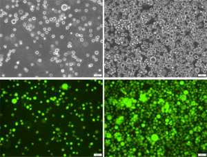 Low and high density cell photos (200x).
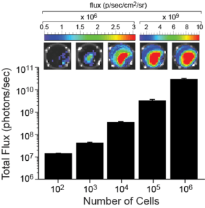 The indicated number of cells were placed in wells of a 96-well plate. After the addition of 3 mg/mL d-luciferin, the plate was immediately imaged using a Xenogen IVIS Spectrum. The total flux (photons/sec) was plotted as a function of cell number.
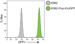 EmGFP Expression
K562-Fluc-EmGFP (green) or parental K562 (grey) cells were fixed with paraformaldehyde and analyzed by flow cytometry.
Complete Growth Medium: Iscove’s Modified Dulbecco’s Medium (IMDM) supplemented with 15% fetal bovine serum (FBS) and 1X Penicillin/Streptomycin.
Grow cells in the indicated medium, subculturing as needed to maintain a density between 4 x 10⁵ and 2 x 10⁶ cells/mL. Passage cells by dilution in fresh medium with occasional complete media changes.
These cells are suitable for in vitro and in vivo experimentation.
The luciferase transgene facilitates noninvasive bioluminescent imaging of implanted cells in vivo and cell-based assays in vitro. eGFP is not recommended for whole animal in-live imaging. Rather, samples can be collected post mortem for analysis by conventional fluorescence microscopy or flow cytometry.
K562 cells form subcutaneous tumors and systemic disseminated tumors at multiple sites (liver, spleen, bone marrow) upon implantation into immunocompromised mice.
These cells can be amplified to generate additional frozen stocks and cryopreservation of low passage stocks in a designated cryopreservation medium is recommended.
These cells were generated by transduction with a self-inactivating Risk Group 3 (RG3) lentiviral vector. All work should be performed under appropriate biosafety conditions per institutional guidelines.

Don’t see what you need in our catalog? Our scientific team engineers cell lines, viral vectors, and assays and provides the characterization data you need to move forward with confidence.