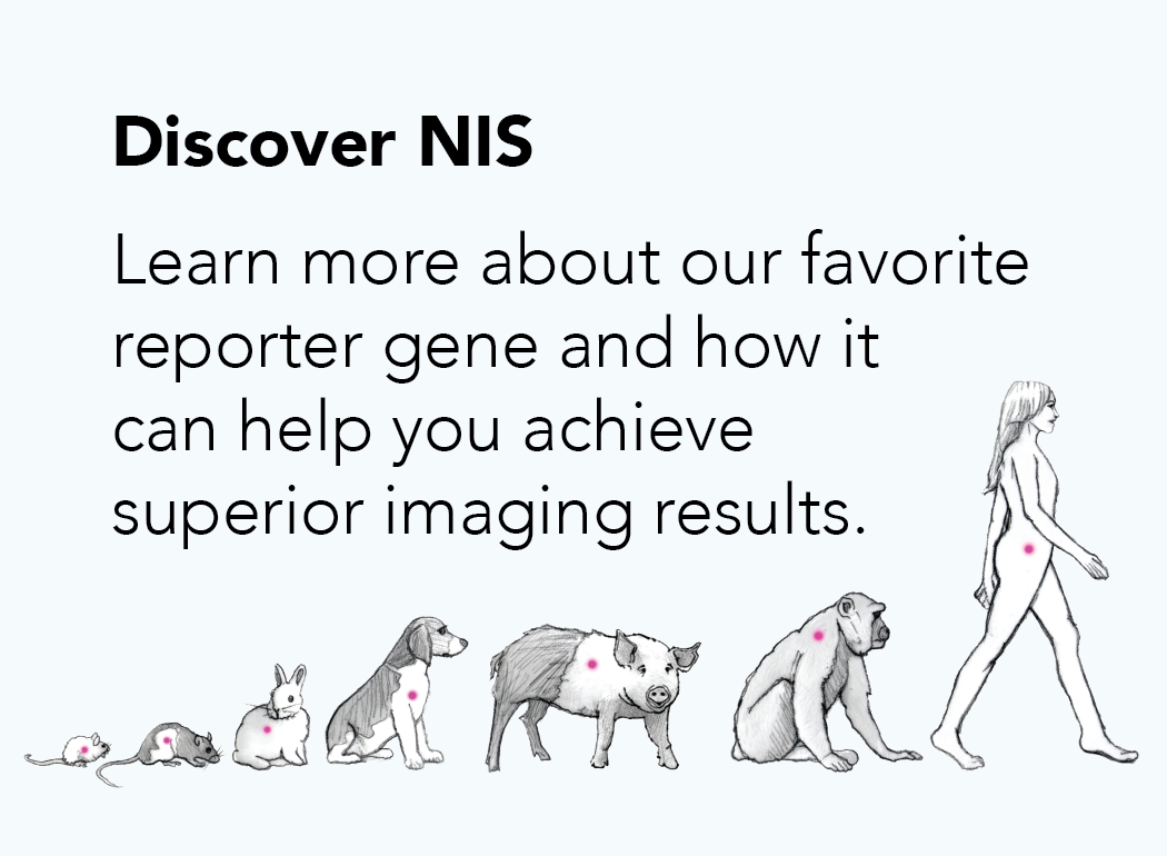
Which Reporter Gene to Use?
So you’ve decided to use noninvasive imaging, but where do you start? With the various reporter options available, how do you know which to choose? When selecting a reporter there are several factors to consider including the type of information you want to get as well as the equipment and resources available to you. Consider the following questions, or check out our reporter comparison chart below.
What degree of sensitivity is required?
Noninvasive imaging reporters have a wide range of sensitivities. If you need to detect as few cells as possible, for example to identify small populations of tumor cells that escape therapy, choose a highly sensitive reporter. In mice and small animals, luciferase (bioluminescence) offers superior sensitivity. However, the bioluminescent signal is attenuated by tissues, so sensitivity decreases with depth of signal and is nonexistent in larger animals. NIS and the other nuclear reporters have excellent sensitivity that is not affected by the depth of signal.
What degree of resolution is required?
Depending on your study, high resolution may be important. Maybe you need to distinguish between different tumor nodules or precisely map the tumor region infected by an oncolytic virus. Intravital microscopy provides the highest resolution, but it is restricted to a very limited region of the animal. Photoacoustic imaging also provides superior resolution but resolution decreases quickly with depth of signal. The
sodium iodide symporter NIS and the other nuclear reporters have excellent 3D resolution that is maintained at any depth of signal. While technological advancements have made tomographic modeling of optical imaging possible, these images are still surface weighted and of relatively low resolution.
Does an immunocompetent model need to be used?
Any immunogenic reporter has the potential to cause tumor rejection in immunocompetent animals. Therefore, if your study requires an immunocompetent model, especially if it is a longitudinal study, a nonimmunogenic reporter is recommended. Fluorescent proteins, iRFP, luciferase, and the nuclear reporter Herpes Simplex Virus Thymidine Kinase (HSVTK) are immunogenic in mammals. The other nuclear reporters and tyrosinase have an advantage in that they are nonimmunogenic if properly species matched; Imanis offers a number of different
species-specific NIS reporter genes. Check out our Science Talk blog
“Betting against Immunology: Choosing the right reporter gene for your oncology models” for a more detailed discussion on how reporter immunogenicity can effect imaging studies.
Do you need quantitative results?
Sometimes having quantitative results are important. Like if you want to compare the efficiency of different gene therapy delivery methods. One of the major advantages of NIS and other nuclear reporters is that they provide fully quantitative results.
What are the target tissues/organs?
Knowing where you will be looking for signal is important for two reasons: reporters have different depths of signal penetration and signal background can be a problem for certain reporters in specific organs. The signal from optical reporters is easily attenuated. Therefore, these reporters aren’t good for deep tissue imaging or imaging in large animals. In contrast, the nuclear reporters have an unlimited depth of signal penetration, making them good for deep tissue imaging and imaging in larger animals and humans. Because most of the nuclear reporters are endogenously expressed in certain tissues in mammals, consideration must be given to the target organ and whether there is expected to be high background in that tissue. Check out our
nuclear reporters page for general guidelines about matching nuclear reporters and target tissues.
Do you want imaging that is translatable to clinical?
Maybe a mouse is all you’re interested in, but maybe you want imaging that’s translatable to the clinic. Optical imaging and photoacoustic imaging of tyrosinase is strictly pre-clinical. Since human variants exist for many of the nuclear reporters and many of the radiotracers are already approved for use in humans, these reporters can be translated to the clinic.
What equipment and reagents are required?
Because imaging equipment is expensive, it is important to consider what imaging equipment is already available at your institution. NIS and other nuclear reporters are imaged with a SPECT or PET machine, and co-imaging with CT or MRI is generally recommended to correlate reporter signal with anatomical location. Luciferase and near-infrared fluorescent protein (iRFP) are imaged with an optical imager equipped with a cooled CCD camera. Other fluorescent proteins (e.g. eGFP, DsRed) can also be imaged using an optical imager, though whole animal imaging is not recommended due to high background autofluorescence. Instead, in-life imaging of fluorescent proteins can be done using an intravital microscope. Post-mortem analysis of fluorescent cells is common and requires either a conventional fluorescence microscope or a flow cytometer. For imaging with iRFP and other fluorescent proteins, it is important to verify that the equipment has the necessary lasers/light sources and filters sets needed for the particular fluorescent protein being used. Tyrosinase imaging requires a photoacoustic imaging system.
Imaging of tyrosinase and fluorescent proteins, including iRFP, does not require additional reagents. Luciferase imaging requires the substrate d-luciferin, which is readily available and relatively inexpensive. Imaging of the nuclear reporters requires an appropriate radiotracer. Depending on your institution, the availability of certain radiotracers may be limited as may be the facilities/training needed to work with specific radioisotopes. Check out our nuclear reporters page, for more information on which radiotracers are used for the different nuclear reporters.
How easy does your imaging need to be?
Some imaging equipment and techniques can be difficult to master. Optical imaging and analysis tends to be easier than nuclear imaging or photoacoustic imaging. Optical imaging also tends to be higher throughput than the other methods, providing more rapid analysis. If your feeling overwhelmed thinking about imaging, the good news is that Imanis can help with your
imaging study or image analysis.
Have you considered multi-modality imaging?
Maybe you want the ultimate sensitivity and the ultimate spatial resolution? Or maybe you just want to confirm your results using a different reporter? Then don’t restrict yourself to a single reporter gene. Using multiple reporters provides the flexibility to image using multiple modalities, so you can, for example, get the sensitivity of luciferase paired with the high resolution tomographic imaging of NIS. Fluorescent proteins can be used to conveniently confirm in vivo imaging results through post-mortem analysis of tissues or samples.
General Conclusions:
Nuclear reporters tend to be the most versatile of the reporters and provide excellent imaging for a wide variety of applications. However, they require very specialized and expensive equipment and have a steeper learning curve than the optical reporters.
Comparison of Reporter Gene Imaging Modalities