CT26.WT (Colorectal Carcinoma)

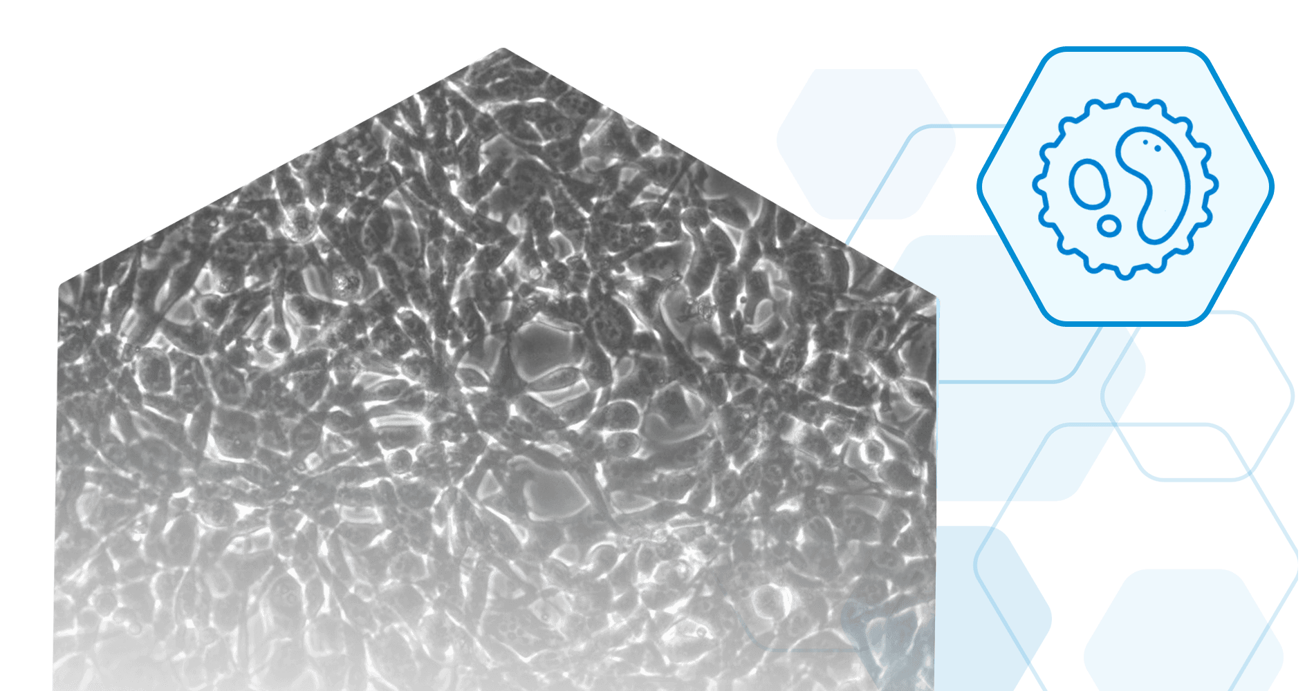
CT26.WT (ATCC® CRL-2638™) is a murine colorectal carcinoma cell line from a BALB/c mouse. It is a clone of the N-nitroso-N-methylrethane-induced undifferentiated CT26 colon carcinoma cell line. The cells are adherent and have a fibroblast morphology. CT26.WT cells will form tumors and metastases post implantation into syngenic BALB/c mice or immunocompromised mice.

*The ATCC trademark and trade name and any and all ATCC catalog numbers are trademarks of the American Type Culture Collection.

Usage Information

CT26.WT cells are suitable for in vitro and in vivo experimentation. Both syngenic BALB/c mice and immunocompromised mice can be used for in life studies, and will form tumors following implantation of the cells. 

The following chart provides some examples of CT26.WT cells used for tumor formation and studies.

Hwang et al. (2021) Journal for ImmunoTherapy of Cancer 9:e002666

Route of ImplantationMiceTumor/MetastasesReferences
IntrasplenicBALB/cLiver metastasesJiao et al. (2014) J Biomed Sci 21: 1 Mi et al. (2006) J Surg Res 131: 97-104
SubcutaneousBALB/cSubcutaneous tumorWang et al. (1995) J Immunol 154: 4685-4692 Hwang et al. (2021) Journal for ImmunoTherapy of Cancer 9:e002666
Tail veinBALB/cLung metastasesWang et al. (1995) J Immunol 154: 4685-4692 Kim et al. (2014) Biochem Pharm 92: 251-265 Gou et al. (2014) PLoS One 9: e85789
IntraperitonealBALB/cPeritoneal tumor metastasesGou et al. (2014) PLoS One 9: e85789 Ryan et al. (2015) Oncogene 34: 1563-1574
Note: The above information is based on available data from the indicated references. It is not meant to be comprehensive and Imanis has not directly tested each condition.

Stable Reporter Cell Lines

Our CT26.WT reporter cell lines can be tracked in vivo, making them great tools for studying the mechanisms of tumor growth and metastasis, as well as evaluating the effects of various drugs or therapies in animals. Our CT26.WT cells are available with a variety of different reporters, including the murine sodium iodide symporter (mNIS), firefly luciferase (Fluc), enhanced green fluorescent protein (eGFP), or near-infrared fluorescent protein (iRFP). Several dual reporter CT26.WT cell lines are available to facilitate multi-modality imaging.

In order to ensure high, constitutive expression of the reporter proteins, our cell lines are generated by lentiviral vector transduction. The lentiviral vectors used for these transductions are self-inactivating (SIN) vectors in which the viral enhancer and promoter has been deleted. This increases the biosafety of the lentiviral vectors by preventing mobilization of replication competent viruses (Miyoshi et al., J Virol. 1998).

Cell & Virus Products Built for Breakthroughs

Don’t see what you need in our catalog? Our scientific team engineers cell lines, viral vectors, and assays and provides as much characterization data as you need to move forward with confidence.