

Near-infrared Fluorescent Protein
For noninvasive in vivo optical imaging
Near infrared-fluorescent protein (iRFP) is an engineered version of the bacteriophytochrome RpBhP2 from the bacteria Rhodopseudomonas palustris. Like other fluorescent proteins, it emits a distinct wavelength of light (peak at 713 nm) after excitation at an appropriate wavelength (peak at 690 nm).
Due to its red-shifted excitation/emission spectrum, iRFP has much lower tissue background than the other fluorescent proteins. Thus, the light emitted by iRFP can be used for noninvasive optical imaging as well as conventional microscopy.
How to use iRFP for imaging
Reagents: iRFP imaging does not require any additional reagents.
Equipment: An optical imager with cooled CCD cameras can be used to detect the light produced by iRFP. The imager must be equipped with an appropriate laser and filter combination (e.g. 675/720 nm) to provide the excitation light and filter the iRFP emitted light. The acquired fluorescence image can be overlayed with a photograph taken by the optical imager to show the relative location of the fluorescent signal.
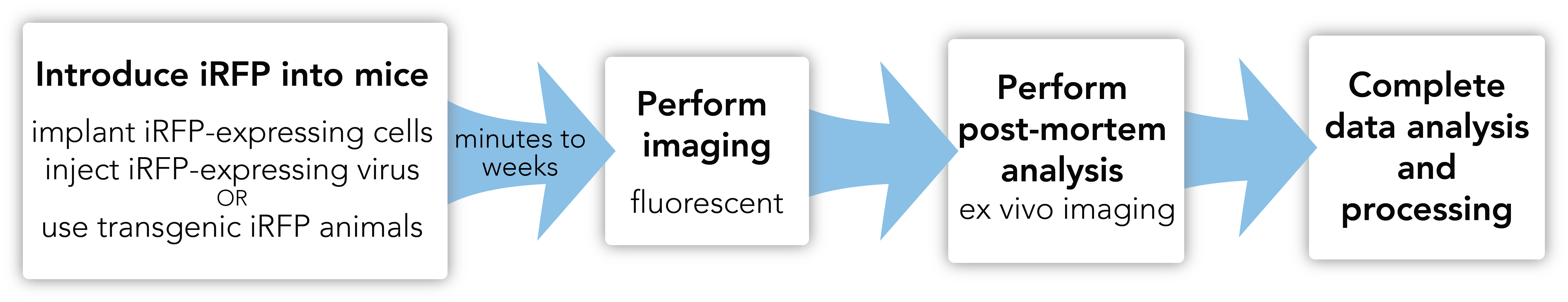
Workflow:
When to use iRFP
Noninvasive fluorescence imaging with iRFP can be used to noninvasively track the biodistribution of cells or viruses in mice and other small animals. Due to its relative ease of use, iRFP is a good reporter option for short-term pre-clinical studies where the signal is relatively close (1-2 cm) to the surface of the animal and high spatial resolution and sensitivity are not required.