Fluorescent proteins are optical reporters that, following excitation by an appropriate wavelength of light, emit light at a specific wavelength. Green fluorescent protein (GFP) is the most commonly used fluorescent reporter. Initially isolated from the jellyfish Aqueoria victoria, GFP has since been engineered to generate the brighter and more photostable enhanced GFP (eGFP), with an excitation wavelength of 488 nm and emission wavelength of 509 nm. Additional mutations of GFP have produced fluorescent proteins emitting different colors of light including yellow, cyan, red (DsRed), and ultraviolet.
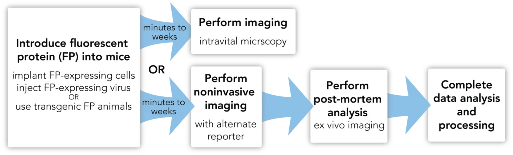
The light emitted by fluorescent proteins can be used to image cells or viruses by conventional or intravital microscopy. Noninvasive optical imaging of fluorescent proteins in mice is hampered by tissue autofluorescence, which makes this type of imaging impractical. Microscopic analyses of fluorescent protein signal in post-mortem samples, however, is frequently used to verify the results of noninvasive imaging using different reporters.




Don’t see what you need in our catalog? Our scientific team engineers cell lines, viral vectors, and assays and provides as much characterization data as you need to move forward with confidence.
