Tyrosinase
For noninvasive in vivo photoacoustic imaging
Tyrosinase (TYR) is an enzyme that converts tyrosine to melanin; the brown pigment associated with skin, hair, and eye color. Since TYR alone is sufficient to trigger melanin production, melanin can be induced in non-melanogenic cells. Once TYR expression is established, melanin production can be noninvasively tracked in vivo by multiple imaging modalities including photoacoustic imaging (PAI), MRI, and PET.
How to use Tyrosinase for imaging
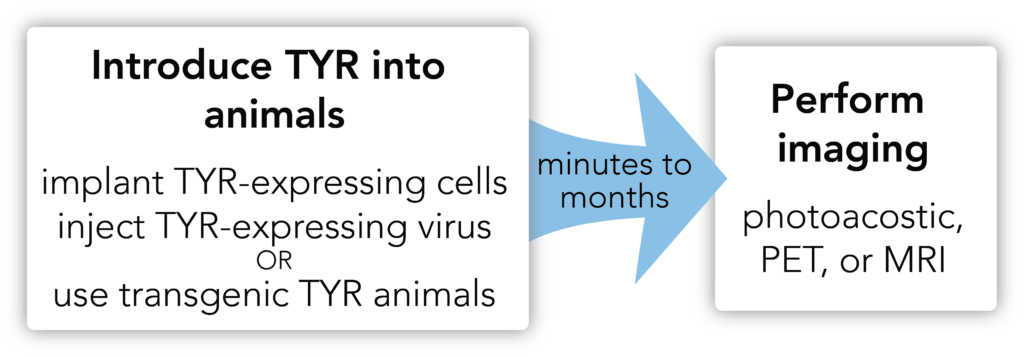
Reagents: No additional reagents are needed for imaging with PAI or MRI. A melanin-avid radiotracer, N-(2-(diethylamino)ethyl)-18F-5-fluoropicolinamide (18F-P3BZA), is required for PET imaging.
Equipment: A photoacoustic imaging machine can be used to detect the melanin produced by TYR at high resolution. PAI is often combined with an ultrasound to indicate the biodistribution of the pigmented proteins. PET machines can also be used to detect melanin-avid radiotracer signal, in combination with CT imaging for anatomical reference.
Workflow:

When to use tyrosinase
Tyrosinase can be used to noninvasively track cells or viruses for pre-clinical gene therapy, oncology, and oncolytic virotherapy studies. Tyrosinase is particularly amenable to photoacoustic imaging (PAI) with high tissue penetration (up to 5 cm), excellent spatial resolution (0.01-1 mm), and relatively high-throughput imaging of soft tissues such as the brain. Currently, PAI is only available for small animal imaging, though large animal imaging technologies are in development.
Imaging considerations
Tyrosinase expression is nonimmunogenic only when species matched, so we recommend using SCID or nude mice for in vivo modeling of hTYR. Although PAI offers high tissue penetration, resolution decreases with increasing tissue depth and dense bone interferes with the light-absorption. Finally, blood hemoglobin also absorbs light during PAI, however imaging at several wavelengths can separate the hemoglobin from the reporter signal. For further reading see Jathoul et al. Nature Photonics. 2015. 22(9):239-46.

