Anti-human NIS SJ1
| Volume | 0.25 ml |
| Antigen | Human sodium iodide symporter (hNIS) |
| Species | Rabbit |
| Reactivity | Human, Rhesus macaque |
| Isotype | IgG |
-
Description
Anti-hNIS antibody SJ1 is a rabbit polyclonal antibody raised against the C-terminal portion (amino acids 468-643) of the human NIS fusion protein (MBP-hNIS).
Western Blot
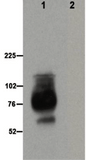
Recommended dilution: 1:3000 to 1:5000
Lane 1: Total membrane protein from Mel624-hNIS-Neo cells (20 µg).
Lane 2: Total membrane protein from control murine CT26.WT-mNIS cells (20 µg). Anti-hNIS Ab was used at 1:2000 dilution. The expected size of hyperglycosylated hNIS is 75-90 kDa (top band) and hypoglycosylated hNIS is 60-65 kDa (bottom band).
For western blotting of NIS proteins, it is recommended that samples be heated at 37°C for 30 minutes prior to loading for SDS-PAGE (do not boil).
Immunofluorescence staining
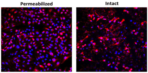 Tissue: Mel624-hNIS-Neo cells
Type: Cytospin of cells on glass slide
Stain: Secondary antibody Alexa 594 with Hoechst 33342
Dilution: 1:500
Recommended dilution: 1:500 to 1:3000
Immunohistochemical staining
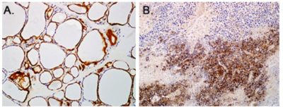 Tissue: (A) human thyroid or (B) VSV-mIFNβ-NIS virus treated mouse tumor xenograft
Type: Paraffin embedded sections
Stain: Anti hNIS SJ1 antibody. Counterstain is hematoxylin.
Dilution: 1:2000
Recommended dilution: 1:2000 to 1:5000
Flow cytometry

Tissue: Human thyroid cell, SW579
Type: Versenized cells
Stain: Anti hNIS SJ1 and anti-rabbit Alexa 594
Dilution: 1:2000
Recommended dilution: 1:2000 to 1:3000
*Note: Users are strongly advised to determine the optimal dilution of antibody to use for their specific applications.
-
Citations
Product Citations
Lakshmanan et al. Thyroid. 2014. 24:878-887.
Knostman et al. BMC Cancer. 2007. 7:137.
Marsee et al. Thyroid. 2005. 15:977-987.
Jhiang et al. J Clin Endocrinol Metab. 2000. 85:2364-2365.
Castro et al. J Endocrinol. 1999. 163:495-504.
Jhiang et al. Endocrinology. 1998. 139:4416-4419.
-
Datasheet/COA
Lot Number REA-IM01 (REA004)