B16F10-Fluc-Neo/eGFP-Puro
| Species | Mouse |
| Cell Type | Melanoma |
| Transgenes | Firefly luciferase (Fluc) Enhanced green fluorescent protein (eGFP) |
| Selection Genes | Neomycin (Neo) Puromycin resistance (Puro) |
-
Description
B16F10-Fluc-Neo/eGFP-Puro is a polyclonal population of the murine melanoma cell line B16F10 (ATCC® CRL-6475™). To achieve stable reporter expression in the polyclonal population, parental B16F10 cells were transduced with LV-Fluc-P2A-Neo (LV011) and LV-eGFP-PGK-Puro (LV031) and selected using G418 and puromycin. LV-Fluc-P2A-Neo encodes the firefly luciferase (Fluc) cDNA linked to the neomycin resistance gene (Neo) via a P2A cleavage peptide under the spleen focus-forming virus (SFFV) promoter. LV-eGFP-PGK-Puro encodes the enhanced green fluorescent protein (eGFP) cDNA under the SFFV promoter and the puromycin resistance gene (Puro) under the phosphoglycerate kinase promoter (PGK).
*The ATCC trademark and trade name and any and all ATCC catalog numbers are trademarks of the American Type Culture Collection.
This cell line has been tested for mycoplasma contamination and is certified mycoplasma free.
The parental B16F10 cell line has been authenticated and certified free of interspecies cross contamination by short tandem repeat (STR) profiling with 9 STR loci.
Due to the immunogenicity of the reporter genes in this cell line, we recommend using immunocompromised mice for in vivo studies.
-
Characterization
Morphology
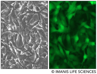 Low and high density cell morphology (200x)
eGFP Expression
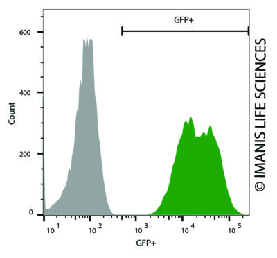 B16F10-Fluc-Neo/eGFP-Puro (green) or control (B16-Fluc-Neo; grey) cells were fixed with paraformaldehyde and analyzed by flow cytometry (20,000 events).
Luciferase Expression
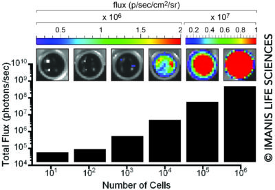 104, 105, or 106 cells were placed in wells of a 96-well plate and 0.3 mg of d-luciferin was added to the indicated wells. The plate was immediately imaged using a Xenogen IVIS Spectrum.
In Vivo Imaging
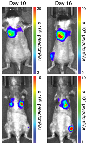 A C57Bl/6 mouse was implanted with 5 x 10⁵ B16F10-Fluc-Neo/eGFP-Puro (CL068) cells through the tail vein. Bioluminescence imaging was performed on the indicated days using an IVIS Spectrum.
 -
Growth Conditions
Complete Growth Medium: DMEM supplemented with 10% FBS, 1X Penicillin/Streptomycin, 0.8 mg/mL G418, and 1 µg/mL puromycin.
The addition of G418 and puromycin to the complete growth medium maintains high dual reporter expression over continued passage of the cells. It is highly recommended, especially if the cells undergo multiple passages prior to being used for studies.
These cells should be grown in the indicated medium and passaged when they reach confluency. For routine passaging, cells are recommended to be split at a 1:10 ratio every 3-4 days.
B16F10 cells produce melanin; accumulation of melanin turns the cells dark brown or black and prolonged melanin secretion turns cell culture medium black. Melanin is toxic and B16F10 cells will die in the presence of excess melanin. Culture medium should be changed as soon as it becomes black, even if the cells are not confluent. Typically, media changes between passages are not required. Different lots of FBS have been observed to affect the rate of melanin production by B16F10 cells.
B16F10 cells trypsinize relatively quickly. Excess trypsinization can damage the cells and care should be taken to quickly neutralize the trypsin upon cell detachment.
-
Usage Information
These cells are suitable for in vitro and in vivo experimentation. B16F10 cells form tumors and pulomary metastases post implantation into syngenic C57BL/6 mice.1 The Fluc transgene facilitates in vivo noninvasive bioluminescent imaging of implanted cells. eGFP is not recommended for whole animal in-live imaging. Rather, samples can be collected postmortem for analysis by conventional fluorescence microscopy or flow cytometry.
The cells can be amplified in vitro and used to generate additional frozen stocks. Cryopreservation of low passage stocks is recommended. Frozen stocks should be preserved in a designated cryopreservation medium.
These cells were generated via lentiviral vector transduction. The lentiviral vector used for transduction was a self-inactivating (SIN) vector in which the viral enhancer and promoter have been deleted. Transcription inactivation of the LTR in the SIN provirus increases biosafety by preventing mobilization by replication competent viruses and enables regulated expression of the genes from the internal promoters without cis-acting effects of the LTR2. Nevertheless, all work with these cells should be performed under biosafety-level 2 (BSL2) conditions by trained personnel. Institutional requirements may permit handling of these cells under BSL1 conditions if certain criteria are met.
References:
1Fidler. Cancer Res. 1975. 35:218-224.
2Miyoshi et al. J Virol 1998. 72:8150-8157. - Datasheet/COA