Daudi-Fluc-EmGFP
| Species | Human |
| Cell Type | B Lymphoblast |
| Transgene | Firefly luciferase (Fluc) Emerald green fluorescent protein (EmGFP) |
-
Description
This is a cell line derived from the human B-lymphoblast Daudi cell line (ATCC® CCL-213TM). Parental Daudi cells were transduced with LV-SFFV-Luc2-P2A-EmGFP (Imanis #LV050) encoding the firefly luciferase (Fluc) cDNA under the spleen focus-forming virus (SFFV) promoter and linked to the emerald green fluorescent protein (EmGFP) via a P2A cleavage peptide. Selection was performed using a methylcellulose-based semi-solid medium. The lentiviral vector is a self-inactivating (SIN) vector in which the viral enhancer and promoter have been deleted.
*The ATCC trademark and trade name and any and all ATCC catalog numbers are trademarks of the American Type Culture Collection.
This cell line has tested negative for mycoplasma contamination.
-
Characterization
Morphology
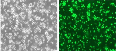 Cell morphology (200x)
Luciferase Expression
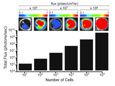
The indicated number of cells were placed in wells of a 96-well plate. After the addition of 3 mg/mL d-luciferin, the plate was immediately imaged using an IVIS Spectrum. The total flux (photons/sec) was plotted as a function of cell number.
Fluorescence Expression
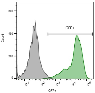
Daudi-Fluc-EmGFP (green) or isotype control (Daudi Parental; grey) cells were fixed with paraformaldehyde and analyzed by flow cytometry.
Surface Receptor Profiling
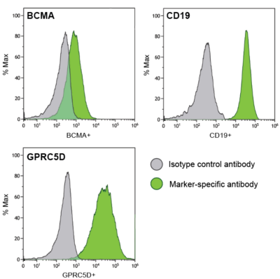
Daudi-Fluc-EmGFP (green) cells were stained with isotype control antibody (grey) or antibody specific to BCMA (top, left), CD19 (top, right), or GPRC5D (bottom) and analyzed by flow cytometry.
In Vivo Growth
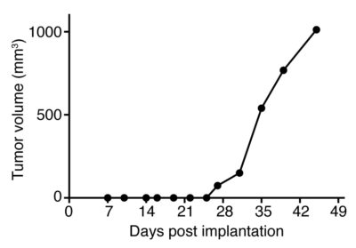
A Nude mouse was implanted with 5 x 10⁶ Daudi-Fluc-EmGFP (CL158) cells in the right hind flank. Tumor growth was monitored over time using calipers.
-
Growth Conditions
Complete Growth Medium: RPMI-1640 Medium (RPMI) containing 10 mM HEPES supplemented with 1 mM sodium pyruvate, 10% fetal bovine serum (FBS), and 1% Penicillin/Streptomycin.
The cells should be subcultured as needed to maintain a density between 5 x 10⁵ and 2 x 10⁶ cells/mL. The cells can be passaged by dilution in fresh complete growth medium. Regular passage using low-speed centrifugation is recommended to limit the amount of debris in cultures.
-
Usage Information
These cells are suitable for in vitro and in vivo experimentation.
The luciferase transgene facilitates in vivo noninvasive bioluminescent imaging of implanted cells. EmGFP is not recommended for whole animal in-live imaging. Rather, samples can be collected post mortem for analysis by conventional fluorescence microscopy or flow cytometry.
These cells were generated via lentiviral vector transduction. The lentiviral vector is a self-inactivating (SIN) vector in which the viral enhancer and promoter have been deleted. Transcription inactivation of the LTR in the SIN provirus increases biosafety by preventing mobilization by replication competent viruses and enables regulated expression of the genes from the internal promoters without cis-acting effects of the LTR1. All work with these cells should be performed under biosafety-level 2 (BSL2) conditions by trained personnel.
-
Datasheet/COA
Lot Number CL-IM222