Anti-Human NIS Antibody (monoclonal, affinity purified)
| Value | 100 ul |
| Antigen | Human sodium iodide symporter (hNIS) |
| Species | Mouse |
| Reactivity | Human |
| Isotype | igG |
-
Description
Monoclonal antibody was raised against peptide NEDLLFFLGQKE LE corresponding to residues 598–621 of the human sodium iodide symporter (hNIS).
Western blotting
 Recommended dilution: 1:1000 to 1:5000
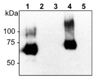
Membrane protein (10 μg; lanes 1-2) or total protein (50 μg; lanes 3-5) was subjected to SDS-PAGE and immunoblotting using monoclonal anti-hNIS antibody (1:3000) and HRP-conjugated anti-mouse secondary antibody (1:5000). Samples: humans NIS (lanes 1 and 4); murine NIS (lanes 2 and 5); negative control lysate (lane 3). Various glycosylated forms of human NIS are detected.
For western blotting of NIS proteins, it is recommended that samples be heated at 37°C for 30 minutes prior to loading for SDS-PAGE (do not boil).
Flow Cytometry
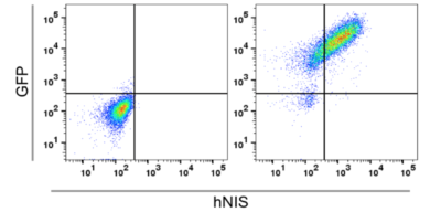
 Tissue: (Left) Parental HeLaH1 cells or (Right) HeLaH1 cells stably expressing hNIS linked to enhanced green fluorescent protein (GFP)
Type: Permeabilized cells
Stain: Anti-NIS Antibody, Alexa Fluor 594-conjugated anti-mouse secondary antibody (1: 1000)
Dilution: 1:500
Recommended Dilution: 1:500
Immunohistochemistry

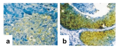
Tissue: Tissue sections were deparafinized. Slides were subjected to antigen retreival using 10% citrate buffer.
a, NIS expression of normal ductal– lobular units in the vicinity of breast cancer assessed with monoclonal antibody against NIS is shown above (magnification, X160).
b, Ductal carcinoma stained with monoclonal antibody against NIS (magnification, x66).
Type: Paraffin Embedded Sections
Stain: Anti hNIS Antibody (REA011)1
Recommended Dilution: 1:5000 to 1: 10,0000
Detection of hNIS by immunohistochemistry was performed by Tazebay et al. 2000.
Note: This antibody recognizes the cytosolic C-terminus of hNIS. Therefore, samples must be permeabilized prior to incubation with anti-human NIS antibody for immunohistochemistry and flow cytometry
- Citations
-
Datasheet/COA
Lot Number REA-IM12