Nalm6-eGFP-Puro
| Species | Human |
| Cell Type | Lymphoma |
| Transgene | Enhanced Green Fluorescent Protein (eGFP) |
| Selection Gene | Puromycin resistance (puro) |
-
Description
Parental Nalm6 cells were transduced with LV-eGFP-PGK-Puro (Imanis #LV031) encoding the enhanced green fluorescent protein (eGFP) cDNA under the spleen focus-forming virus (SFFV) promoter and the puromycin resistance gene (Puro) under the phosphoglycerate kinase (PGK) promoter. A high eGFP expressing population was generated by selection using puromycin followed by selection using a methylcellulose based semi-solid medium.
This cell line has been tested for mycoplasma contamination and is certified mycoplasma free.
The parental Nalm6 cell line was authenticated and certified free of interspecies cross contamination by STR profiling.
-
Reporter Validation
Morphology
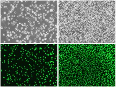
Low and high density cell morphology (200x)
eGFP Expression
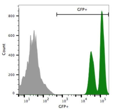
Nalm6-eGFP-Puro (green) or isotype control (Nalm6-Fluc-Puro; grey) cells were fixed with paraformaldehyde and analyzed by flow cytometry.
-
Growth Conditions
Complete Growth Medium: RPMI-1640 Medium (RPMI) containing 10mM HEPES supplemented with 10% FBS, 1% Penicillin/Streptomycin, and 1 µg/mL puromycin.
The addition of puromycin to the complete growth medium maintains high reporter expression over continued passage of the cells. It is recommended, especially if the cells undergo multiple passages prior to being used for studies.
These cells should be grown in the indicated medium and subcultured as needed to maintain a density between 3 x 10⁵ and 3 x 10⁶ cells/mL. For optimal growth, frequent passaging at lower subcultivation ratios is recommended. The cells can be passaged by dilution in fresh medium. Regular passaging using centrifugation is recommended to limit the amount of debris in cultures.
-
Usage Information
These cells are suitable for in vitro and in vivo experimentation.
Nalm6 cells form tumors and metastases at multiple sites, including the liver, lymph nodes, spleen, long bones, periodontal region, and bone marrow, upon implantation into immuncompromised mice1,2.
eGFP is not recommended for whole animal in-live imaging. Rather, samples can be collected post mortem for analysis by conventional fluorescence microscopy or flow cytometry.
These cells were generated via lentiviral vector transduction. The lentiviral vector used for transduction was a self-inactivating (SIN) vector in which the viral enhancer and promoter have been deleted. Transcription inactivation of the LTR in the SIN provirus increases biosafety by preventing mobilization by replication competent viruses and enables regulated expression of the genes from the internal promoters without cis-acting effects of the LTR2. Nevertheless, all work with these cells should be performed under biosafety-level 2 (BSL2) conditions by trained personnel. Institutional requirements may permit handling of these cells under BSL1 conditions if certain criteria are met.
-
Datasheet/COA
Lot Number CL-IM149