VV(Li)-iCBG-RFP-GusA
| Strain | Lister (Li) |
| Transgenes | Tet-repressor (tetR) Red fluorescence protein (RFP) Click beetle green luciferase (CBG) β-glucuronidase (GusA) |
| Titer | >3e6 TCID50 units/mL (see description) |
| Risk Group | 2 |
-
Description
VV(Li)-iCBG-RFP-GusA is a recombinant vaccinia virus (VV) encoding the following additional transcription units: 1) the tet-repressor (tetR) cDNA under transcriptional control of the VV synthetic early late (pSEL) promoter, 2) the tet-operator sequence tetO followed by the click beetle green luciferase (CBG)-red fluorescence protein (RFP) fusion cDNA under transcriptional control of the VV synthetic late (pSL) promoter, and 3) the β-glucuronidase (GusA) cDNA under transcriptional control of the p11 promoter. These additional transcription units are inserted into, and disrupt the function of, the viral F14.5L, J2R (thymidine kinase), and A56R (hemagglutinin) genes, respectively. Expression of the CBG-RFP fusion protein is under the control of an inducible promoter-system, whereby addition of doxycycline induces the expression of CBG-RFP fusion protein (see diagram below). The purchased virus is ready for use in cell-killing assays or can be amplified to create additional virus stocks.

The virus titer is functional infectious virus, not total virus particles, in clarified supernatant. The titer is determined using an end-point dilution assay that measures the amount of virus required to produce cytopathic effects in 50% of infected cells (tissue culture infective dose per mL).
Note: A USDA permit is required for shipping Vaccinia Virus. Click here to fill out Application Form VS 16-3 to obtain a USDA APHIS VS 16-6 or 16-6A permit.
-
Propagation
This virus should be handled in a BSL2 facility by trained personnel following proper biocontainment practices.
Basic protocol
(All volumes are given for a T75 flask; increase or decrease as needed.)
1. Seed producer cells (e.g. Vero) in complete medium at an appropriate density to achieve 80-90% confluency at the time of infection and incubate in an appropriate incubator.
2. Thaw the virus stock on ice.
3. In a microcentrifuge tube, prepare virus at a MOI of 0.1 in 3 mL total serum free media.
4. Remove culture medium from cells and replace with prepared virus. Return cells to incubator.
5. After 2-3 hours, remove virus innoculum and replace with 12 mL complete medium. Return cells to incubator.
6. Harvest supernatant at 48 hours post-infection.
7. Freeze-thaw or sonicate the supernatant and cells for 3 cycles.
8. Clarify the supernatant by low-speed centrifugation and purify through a sucrose cushion.
9. Resuspend the viral pellet in 1mM TrisCl, pH 9. Determine virus titer using appropriate methods. Store at -80C. -
Transgene Validation
Infectivity
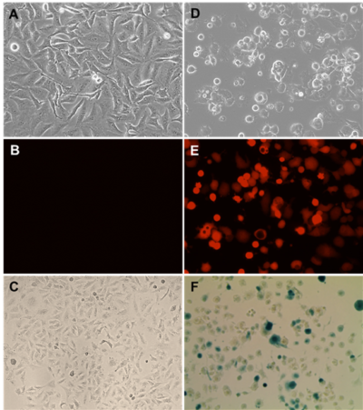
(A-C): Mock-infected A549 cells.
(D-F): VV(Li)-iCBG-RFP-GusA infected A549 cells (MOI 0.1) in the presence of 1µg/ml doxycycline at 48 h.p.i.
(C&F): For β-glucuronidase activity, cells were incubated with media containing X-Gluc (5-bromo-4-chloro-3-indolyl-beta-glucuronide).
Luciferase Expression

Vero cells were mock-infected or infected with VV(Li)-iCBG-RFP-GusA (MOI of 0.1) in the presence (+Dox) or absence (-Dox) of 1µg/ml doxycycline. After 48 hours Click beetle luciferase substrate d-luciferin was added to the wells and luminescence (RLU) was immediately measured using a microplate reader.
-
Datasheet/COA
Lot Number OV-IM31