

Stable Cell Lines
Imanis offers stable cell lines to support in vitro and in vivo oncology and immune-oncology applications. Our constitutive Reporter Cell Lines contain luciferase, fluorescent proteins, or the NIS reporter and can be used in vitro or to monitor tumors in living animals. Our Tumor Antigen Cell Lines have been engineered to express altered levels of common tummor associated antigens and therapeutic targets, like CD19, EGFR, and Her2.
Human Xenograft Models
- RPMI-8226 (Multiple Myeloma)
- U266B1 (Multiple Myeloma)
- Raji (B Lymphoblast)
- Daudi (B Lymphoblast)
- K562 (Chronic Myelogenous Leukemia)
- Nalm6 (Leukemia)
- A549 (Lung Adenocarcinoma)
- HT1080 (Fibrosarcoma)
- HCT116 (Colorectal Carcinoma)
- A375 (Melanoma)
- PC3 (Prostate)
Mouse Syngeneic Models
Higher Reporter Gene Expression
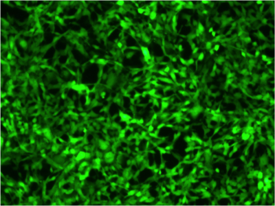
Imanis reporter gene cell lines have bright constitutive reporter expression for more sensitive detection of cells in vitro and in vivo. Delivered by lentiviral vectors, the reporter genes are stably integrated into the cell genome. Reporter expression is driven by the constitutively active SFFV promoter, which delivers high, continued reporter expression in cell cultures and tumors. Xenograph and sygeneic tumor models are available with one, or a combination, of our four main reporters: enhanced green fluorescent protein (eGFP), firefly luciferase (Fluc), near-infrared fluorescent protein (iRFP), or the sodium-iodide symporter (NIS). These reporters offer the flexibility to choose between imaging modalities and equipment, whether it be intravital or conventional microscopy using eGFP, noninvasive in-life imaging using Fluc or iRFP, or noninvasive, high-resolution 3D PET/SPECT imaging using NIS.
NEWTumor Models for Therapeutic Development
Imanis tumor antigen cell lines express altered levels of tumor associate antigens for evaluating the efficacy of targeted cancer therapies. Each cell line is characterized for expression of other relevant tumor associated antigens, to facilitate studies using tandem or dual therapeutic target strategies. Panels provide a range of target expression levels to assess therapeutic efficacy as related to target receptor density. Most of our cell lines also contain a luciferase reporter gene to facilitate both in vitro and in vivo efficacy studies. We are actively developing additional tumor antigen cell lines to add to our catalog. Contact us to learn more about what’s in our current pipeline and provide feedback on research needs.